The proposed framework consists of three main parts:
Part I: Cortical surface reconstruction and atlas construction for population-level healthy joint modeling
Part II: Elastic shape model to estimate healthy cortical surface from diseased joints
Part III: Quantitative filtration and visualization of bone proliferation regions
The complete workflow is illustrated in Figure 1 (cortical reconstruction → atlas-based shape analysis → proliferation detection and visualization).
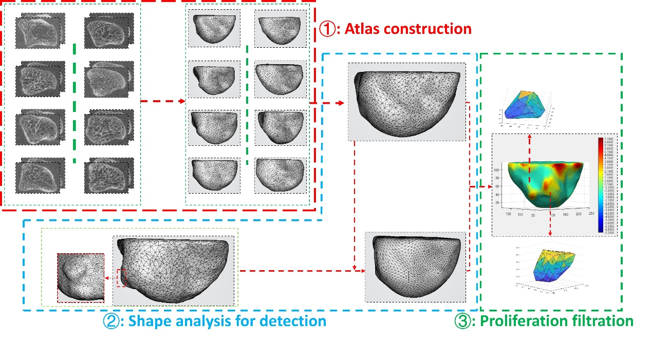
Figure 1. The workflow of bone proliferation detection.
The first step (in the red dashed pane) constructs atlas for population-level analysis.
The second step (in the blue dashed pane) detects joint proliferation using shape analysis methods.
The third step (in the green dashed pane) filtrates proliferation regions (best viewed in color).
Part I: Atlas Construction
Based on previous cortical surface reconstruction work, enhanced with 3D nnU-Net segmentation and level-set refinement using GPU AutoDiff acceleration
nnU-Net trained on physician-annotated joint volume masks from CUHK
Offline atlas (canonical healthy joint surface) constructed using Karcher mean estimation under elastic shape distance
Displacement fields parameterized by SIREN neural network
Part II: Shape Analysis-based Joint Proliferation Detection
Core technique: Elastic deformation model with neural displacement field parameterization (SIREN network).
Elastic distance defined with multiple energy terms (default parameters: 200, 200, 100, 20)
Optimized using PyTorch + Adam + ReduceLROnPlateau scheduler (initial lr = 1e-4)
Atlas estimation formulated as Karcher mean problem with rigid alignment preprocessing
Algorithm 1 describes the iterative random sampling and displacement field update procedure
Part III: Quantitative Proliferation Detection
Rigid alignment of diseased surface to atlas → compute displacement field → deform atlas